|
RETINAL EXAMINATION and DIGITAL RETINAL PHOTOGRAPHY

Yang Low
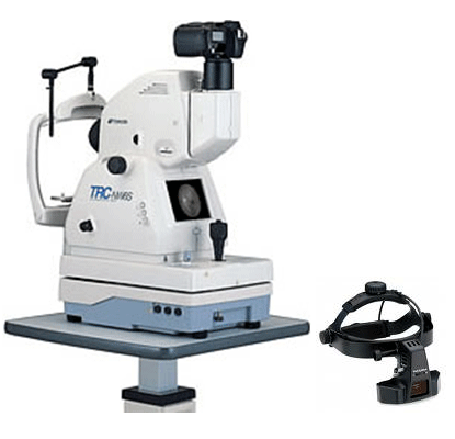
Ophthalmic Opticians is the first to introduce retinal photography to
Miri back in 1997, since then we have upgraded our camera from a
Polaroid one used extensively at the time, to the present more
advanced digital retinal camera.
At present, we are still the only private optometrist to have this
service in Miri.
We pioneered, Diabetic Retinopathy Screening in Miri with our
community pilot projects to established the prevalence of diabetic
retinopathy in Miri.
What is
Retinal Photography
Retinal
Photography takes a high resolution digital photograph of the retina.
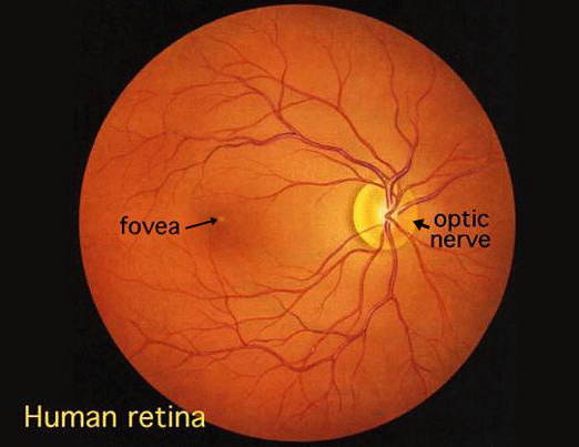
The retina is the light sensitive inner lining of the eye. It's made
of several individual layers and acts in a similar way to the film in
a traditional camera. Light enters the eye, and is focused by the
cornea (the front of the eye) and intra-ocular lens (the lens within
the eye) on to the retina. This information is then transmitted to the
brain via the optic nerve.
The
photograph allows us to view all the blood vessels around the retina,
the macula (the most sensitive part of the retina) and the optic
nerve. In fact, this is the only non- invasive way blood vessels in
our body can be viewed.

What does the test involve?
This test
is very quick and painless. It involves sitting in front of a camera
and fixating on a set target. Within a single flash a digital
photograph of the retina is taken.
Why is it important?
Over time,
there are many conditions that can directly affect our eyes and
bodies. Since we can get a direct view of the blood vessels in the
eye, we can identify these conditions more accurately with a picture.
Conditions
that can effect our sight include:
• Diabetes
• High blood pressure
• High cholesterol levels
• Retinal holes and retinal detachment.
• Glaucoma
• Age-related macular degeneration
Since, the
eye is the only place in the body where the internal blood vessels can
be viewed directly and we are looking for any abnormalities or changes
that may have occurred, we highly recommend this to be done as a
routine checkup. By taking a digital image we can enlarge different
sections of the retina to get a more detailed view so even the
smallest changes to structures can be monitored and managed more
effectively. Over time we will be able to pull forward previous images
to compare any changes.
Why should I have retinal photography?
The
benefits of the test are seen in the future rather than immediately.
So we recommend this test as part of every eye health consultation.
Put simply, this simple & inexpensive test may prove to SAVE YOUR
SIGHT.
Indirect Ophthalmoscopy

Ophthalmoscopy or Funduscopy is an examination of the back part of the
eyeball (fundus), which includes the retina, optic disc, choroid, and
blood vessels.
How the test is performed
There are different types of ophthalmoscopy.
• Direct ophthalmoscopy
• Indirect ophthalmoscopy
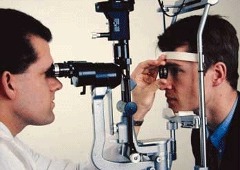
• Slit-lamp ophthalmoscopy
Direct ophthalmoscopy: You will be seated in a darkened room. The
health care provider performs this common exam by shining a beam of
light through the pupil using an instrument called an ophthalmoscope.
An ophthalmoscope is about the size of a flashlight. It allows the
examiner to view the back of the eyeball.
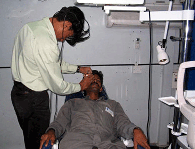
Indirect ophthalmoscopy: You will either lie or sit in a semi-reclined
position. The health care provider holds your eye open while shining a
very bright light into the eye using an instrument worn on the head.
(The instrument looks like a miner's light.) Some pressure may be
applied to the eyeball using a small, blunt tool. You will be asked to
look in various directions.
For information on slit-lamp ophthalmoscopy, see the article:
Slit-lamp exam
Opthalmoscopy examination takes about 5 and 10 minutes.
How to prepare for the test
Indirect ophthalmoscopy and slit-lamp ophthalmoscopy are performed
after eye drops are placed to widen (dilate) the pupils. Direct
ophthalmoscopy can be performed with or without dilation of the pupil.
The eye drops may make it hard to focus your eyes for several hours.
You should arrange to have someone else to drive after the
examination. Wearing sunglasses or tinted lenses will help make your
dilated pupils more comfortable.
You should tell the health care provider if you:
• Are allergic to any medications
• Are taking any medications
• Have glaucoma or a family history of glaucoma

How the test will feel
The bright light will be uncomfortable, but the test is not painful.
You may have a brief sensation of seeing images after the light shines
in your eyes. The light is brighter with indirect ophthalmoscopy, so
the sensation of seeing after-images may be greater.
Pressure put on the eyeball during indirect ophthalmoscopy by may be
slightly uncomfortable, but should not be painful.
If eyedrops are used, they may produce a brief stinging sensation when
placed in the eyes and an unusual taste in the mouth.
Why the test is performed
Ophthalmoscopy is done as part of a routine physical or comprehensive
eye examination.
It is used to detect and evaluate symptoms of retinal detachment or
eye diseases such as glaucoma.
Ophthalmoscopy may also be done if you have signs or symptoms of high
blood pressure, diabetes, or other diseases that affect the blood
vessels.
What abnormal results mean
Abnormal results may be due to eye diseases such as a detached retina,
optic nerve problems, macular degeneration, and changes caused by
glaucoma. High blood pressure can also be detected.
Additional conditions under which the test may be performed:
• CMV retinitis
• Diabetes
• Hypertensive retinopathy
• Macular degeneration
• Melanoma of the eye
What the risks are
The test itself involves no risk. The dilating eye drops may rarely
produce nausea, vomiting, dryness of the mouth, flushing, dizziness,
or an attack of narrow-angle glaucoma. If the latter is suspected,
drops generally are not used.
Special considerations
Ophthalmoscopy is considered to be 90-95% accurate and can detect the
early stages and effects of many serious diseases. |