Viewing the retina
 Back in 1986 When I conducted my full eye examination the only practical way to examine the back of an eye was to use a direct ophthalmoscope and the view I got, even with an extremely good ophthalmoscope, is very limited.
Consequently, I got a very good indirect ophthalmoscope to get a better 3 dimensional view but I had to spend quite a lot of time looking very carefully into each eye and it’s a very skilled task simply being able to see what is there before I can start to work out if anything is going wrong.
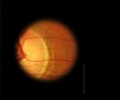
This is what the back of an eye looks like through an ophthalmoscope – you can’t see very much at a time so I had to move around a lot and piece together the different small images in my mind.
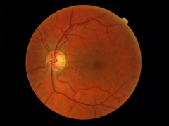 In 1995, I bought our first Polaroid Fundus Camera. Fundus Cameras take images of the back of an eye (the fundus oculi) and we were the first Optometrist to start a Retinal Photo Diabetic Retinopathy Screening Service in Malaysia. We were able to publish our findings with recommendations that the Malaysian Ministry of Health start this service Nationwide.
In 2005, we upgraded our fundus camera to a digital camera.
The advantage of a camera over an ophthalmoscope is that we now have a permanent record to compare images of retinal disease progression. I can also see a much wider area of the back of the eye at any one time. This is what the same eye looks like using a fundus camera: Our polaroid camera at that time had a 45° view while our digital camera had a 80° view of the inside of the eye.
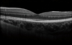 2014 and we are proud to announce the arrival of our NEW Topcon 3D OCT – 1 Maestro. My view of the retina now is to the resolution of microns .….. and in 3 dimension.
An Optical Coherence Tomography Scan is equivalent to a MRI scan for the eye.
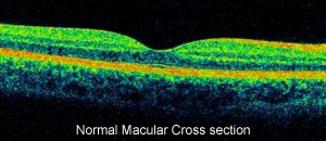
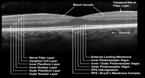
Our OCT camera not only takes better, more detailed images but also can take 50,000 A scans per second of the front and back of the eye which are effectively cross-sections through the cornea iris and retina. I can show up these structures of the eye in incredibly fine detail. This particular scan is through the macula shows very clearly how the nerve fibres at the top are angled away from the very central part to allow more light through to the photo-receptors underneath. The overall thickness of the whole retina shown on this scan is just over half a millimetre. It can then put all the scans together to produce a three dimensional image of the retina which allows me to see and understand much better the very fine changes which can be the indications of any abnormalities.
The Topcon 3D OCT – 1 Maestro Optical
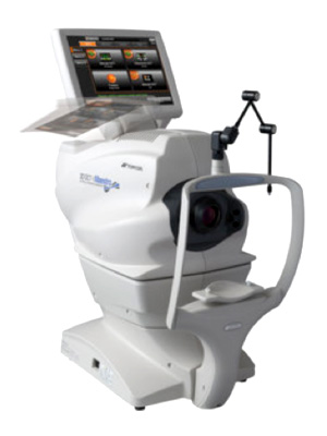
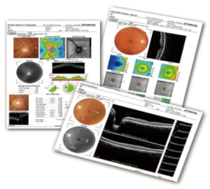
Coherence Tomographer
This state of the art equipment not only takes high resolution retinal photographs but can also performs extremely high resolution scans of the back of the eye enabling early detection of many eye disorders, in particular glaucoma and macular degeneration.
OCT and the normal Eye
Ocular Coherence Tomography (OCT) is an imaging test that allows for very fine detailed visualisation of the optic nerve head and retina.
OCT & Glaucoma
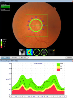
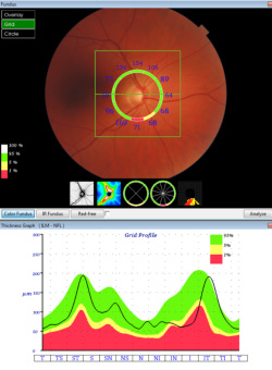
The damage glaucoma causes to the optic nerve can be detected in two general ways: either with the way the nerve is working (i.e. problems with vision) which traditional has to be subjectively acquired or by observing objectively for any abnormal changes in the structure of the nerve.
Problems with the appearance of the optic nerve can be seen long before people have any trouble with their vision. Detecting these problems with the structure of the optic nerve is therefore very important as an early sign of glaucoma (before subjective reduction in vision is noticed) as well as that glaucoma may be getting worse.
There are a number of different ways of recording the appearance of the optic nerve. With the advent of high quality eye cameras it became possible to record the optic nerve in life like detail and optic nerve photos remain a very useful way of imaging in glaucoma. By comparing new and old photographs it is possible to see changes in the nerve, although it can be difficult to get exact measurements of the change. OCT gives a new way of recording and measuring the structure of the optic nerve in great detail.
The OCT machine is similar to ultrasound but rather than using sound waves to create an image it uses light.
This can tell us a great deal about the optic nerve. Each section of the nerve can be measured and compared to the general population to see if there is anything obviously abnormal. The optic nerve is usually less than 2mm in diameter and OCT provides measurements of the optic nerve shape and thickness with a resolution of about 6 microns (1 micron=1/1000th of a mm). In many cases this can help make the diagnosis of glaucoma a lot easier.
The real strength of OCT however is in its ability to detect changes over time.
When the OCT is repeated after a period of time the new scans can be compared to the old ones, allowing detection of changes which may be too subtle to have been noticed with older technologies, such as colour photos or drawings.
Having an OCT picture taken is very similar to having a normal picture taken of your eye. It is not generally required to touch the eye but some people may need to have eye drops put in to dilate the pupil in order to get the best pictures. The scan itself takes only a few seconds.
Anterior Segment OCT can also be done to evaluate the drainage capabilities of the eye, which is also an important factor in the management of Glaucoma patients.
OCT & Macular Degeneration
Your retina is the light-sensing layer at the back of your eye. Like the film in a camera, it picks up images which are transmitted to your brain via your optic nerve. Your brain does the real work of seeing, making sense of all those images. Any damage or deterioration in your eye or optic nerve that reduces the quality of the images your brain gets can affect your ability to see.
The macula enables us to see detail, so any damage to it affects our ability to read or do anything that requires precise vision, such as driving.
Macula Degeneration (MD), is also know as Age-related macula degeneration (AMD or ARMD)
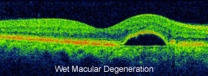
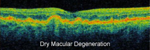
There are two types of MD - known as Dry MD and Wet MD. The Dry form results in a gradual loss of central vision. The Wet form is characterised by a sudden loss of vision and is caused by abnormal blood vessels growing into the retina. Symptoms may include distortion, where straight lines appear wavy or bent or difficulty in reading or other activities that require fine vision.
MD is thought to be caused by genetic and environmental factors. People over the age of 50 are particularly at risk. If you smoke or have a family history of MD, your risk of developing the disease is much greater.
The early detection of any form of MD is crucial because the earlier you seek treatment, the more likely you are to have a better outcome compared to those who wait. Early detection of changes can allow you to take steps to slow the progression of MD.
The OCT machine is similar to ultrasound but rather than using sound waves to create an image it uses light. The OCT takes high resolution cross-sectional images of the macula. By comparing the structure and thickness of the layers measured by the OCT against a normal healthy retina, your Optometrist can detect MD even at a very early stage.
When the OCT is repeated after a period of time the new scans can be compared to the old ones, allowing detection of changes which may be too subtle to have been noticed with older technologies, such as colour photos or drawings.
Treatment options are dependent on the stage and type of the disease. Current treatments aim to keep the best vision for as long as possible and in some cases may potentially provide visual improvement, but there is presently no cure. Early detection is vital in saving sight.
